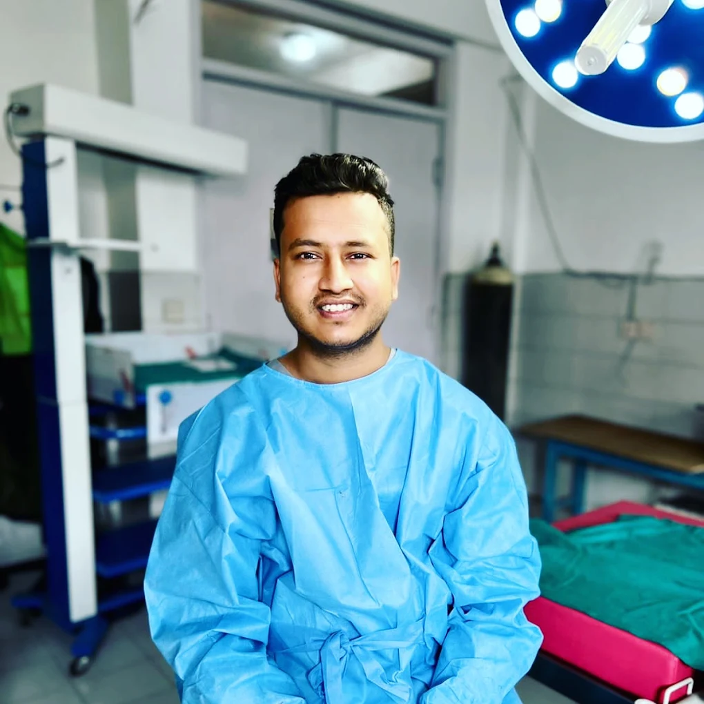
Dr. Rajan Shah: Consultant Laparoscopic Gynecologist & Infertility Specialist in Kathmandu, Nepal
Published on August 14, 2026
Dr. Rajan Shah’s official clinical practice provides advanced minimally invasive gynecological surgery and comprehensive reproductive medicine in Kathmandu, Nepal. As a Consultant Obstetrician and Gynecologist (NMC No. 16365) practicing at Everest Hospital, New Baneshwor, Dr. Shah specializes in laparoscopic management of complex pelvic conditions including uterine fibroids, ovarian cysts, deep endometriosis, adenomyosis, and ectopic pregnancies alongside structured infertility evaluations. Having performed more than 5,500 gynecological laparoscopic surgeries, his practice integrates high-definition surgical precision with fertility-preserving techniques to support long-term women’s health.
1. Professional Background and Medical Credentials
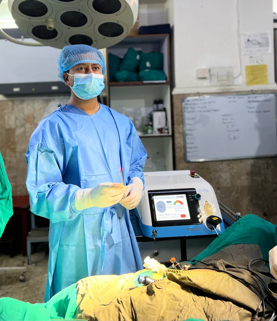
Safe surgical and reproductive care depends entirely on rigorous, accredited medical training and ongoing surgical skill development.
Dr. Shah completed his undergraduate medical education (MBBS) through Tribhuvan University, followed by his Master’s degree in Obstetrics and Gynecology from the B.P. Koirala Institute of Health Sciences (BPKIHS), a premier tertiary health institution in Nepal.
To refine his operative precision in minimally invasive techniques, he pursued specialized post-graduate surgical training, earning a Fellowship in Minimal Access Surgery (F.MAS, awarded with Merit) and a Diploma in Minimal Access Surgery (D.MAS). You can read more about his detailed surgical training and credentials and active involvement in national continuing medical education programs.
Dr. Shah maintains active registration with the Nepal Medical Council (NMC No. 16365) and dedicates his full-time clinical hours to patient care, operating, and reproductive medicine consultations at Everest Hospital Pvt. Ltd., located in New Baneshwor, Kathmandu.
2. Advanced Gynecological Laparoscopy: Surgical Expertise
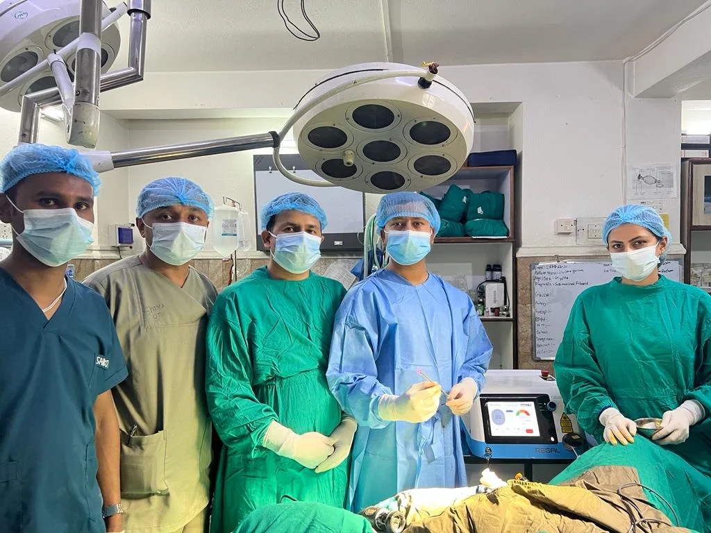
Gynecological laparoscopy is a surgical discipline that uses small keyhole incisions (typically 5 mm to 10 mm) rather than a large abdominal incision. Through an illuminated optical system and specialized microsurgical instruments, pelvic pathology can be diagnosed and treated with minimal tissue disruption.
Dr. Shah has completed over 5,500 laparoscopic procedures across a wide spectrum of complex gynecological conditions.
Uterine Fibroid Surgery (Laparoscopic Myomectomy)
Uterine leiomyomas (fibroids) are non-cancerous muscular tumors that frequently lead to abnormal uterine bleeding, pelvic pressure, frequent urination, and reproductive complications. Dr. Shah performs laparoscopic myomectomy to excise fibroids while meticulously reconstructing the uterine myometrial layers in multiple planes. This layered reconstruction preserves structural integrity for future pregnancies.
Endometriosis and Adenomyosis Management
Endometriosis occurs when tissue resembling the lining of the uterus grows outside the uterine cavity, causing severe pelvic pain, chronic inflammation, and subfertility. Dr. Shah applies precise laparoscopic excision techniques to eliminate peritoneal deposits, endometriomas (chocolate cysts), and deep pelvic adhesions while protecting vital adjacent structures like the ureters, bladder, and bowel.
Ovarian Cyst Surgery (Laparoscopic Cystectomy)
Benign ovarian neoplasms, including dermoid cysts, serous/mucinous cystadenomas, and endometriomas, require precise surgical removal. Using fine dissection plane separation, Dr. Shah enucleates the cyst wall completely while preserving the healthy adjacent ovarian cortex and ovarian reserve.
Laparoscopic Hysterectomy
When conservative medical options have been exhausted or when definitive management is indicated for conditions such as severe adenomyosis, large multi-fibroid uteri, or persistent abnormal uterine bleeding, Total Laparoscopic Hysterectomy (TLH) is performed through minimal access ports. This significantly lowers blood loss, reduces postoperative discomfort, and avoids open abdominal scarring.
For a full breakdown of conditions treated, explore our minimally invasive gynecological procedures.
3. Cutting-Edge Operative Technology: 3D 4K RUBINA with ICG
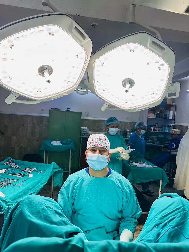
Surgical safety and precision rely heavily on visualization quality. Dr. Rajan Shah operates using the Karl Storz 3D 4K RUBINA with Indocyanine Green (ICG) fluorescence imaging system, which offers several distinct clinical advantages:
-
Unrivaled Anatomical Clarity: Native 4K imaging magnifies the pelvic anatomy on medical monitors, allowing clear distinction between pathological lesions and healthy organs.
-
True Spatial Depth Perception: Conventional 2D laparoscopy flattens the operative field, requiring mental depth compensation. Stereoscopic 3D visualization improves manual dexterity during delicate tasks such as intracorporeal suturing of the uterine wall during a myomectomy.
-
Fluorescence Vascular Assessment: Near-infrared light combined with intravenous Indocyanine Green (ICG) dye helps assess blood perfusion across reconstructed uterine tissue and identify tissue planes around delicate pelvic retroperitoneal spaces.
4. Uterus- and Ovary-Preserving Surgery
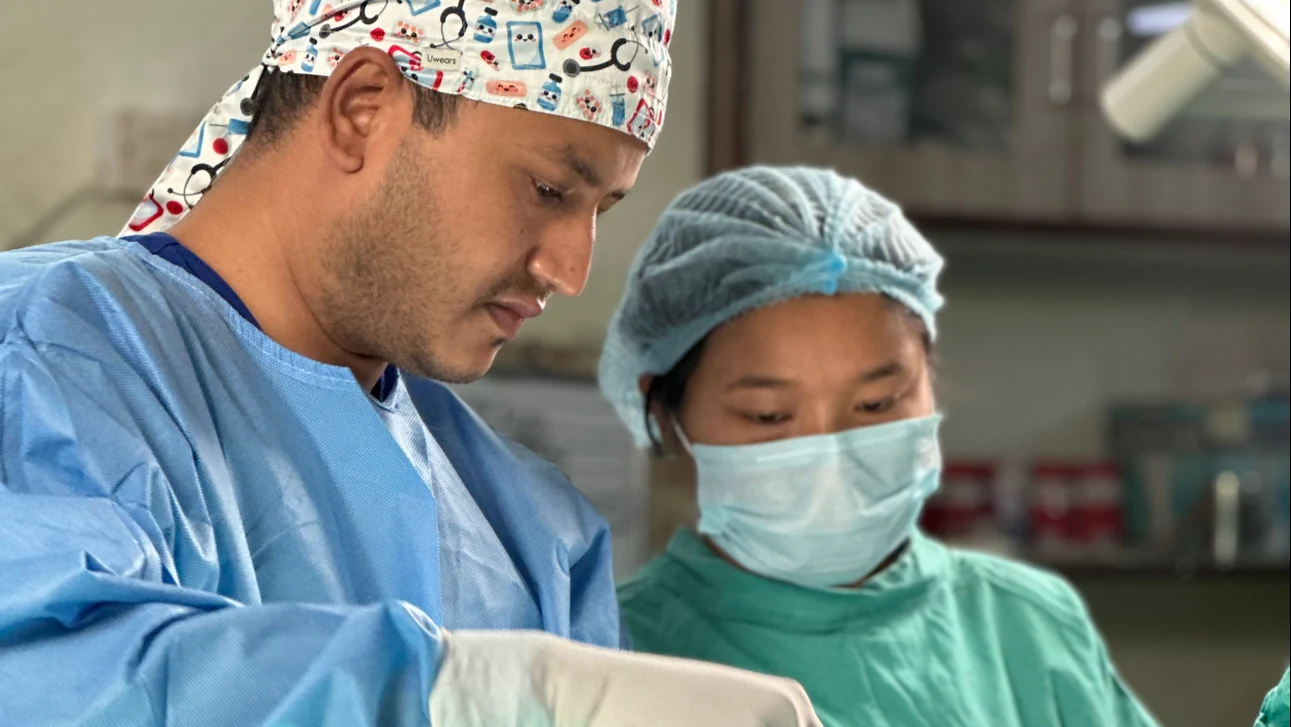
For women in their reproductive years, preserving functional anatomy is often an essential consideration alongside symptom relief.
A central focus of Dr. Shah’s surgical practice is organ preservation:
-
Meticulous Myometrial Suture Technique: In laparoscopic myomectomy, the uterine wall defect is repaired using multi-layered, tension-controlled continuous suturing. This promotes solid tissue healing and supports the uterine wall during future pregnancies.
-
Ovarian Reserve Protection: During ovarian cystectomy, gentle mechanical traction and targeted bipolar/cold-scissor dissection are prioritized over excessive thermal energy. Minimizing electrocoagulation near the ovarian hilum protects surrounding primordial follicles and helps maintain the patient’s Anti-Müllerian Hormone (AMH) levels.
-
Adhesiolysis for Tubo-Ovarian Mobility: Pelvic adhesions from previous infections or endometriosis can tether the fallopian tubes and ovaries, impairing natural ovum pick-up. Restoring normal pelvic anatomy helps re-establish natural fertility pathways.
5. Comprehensive Infertility Evaluation and Reproductive Medicine
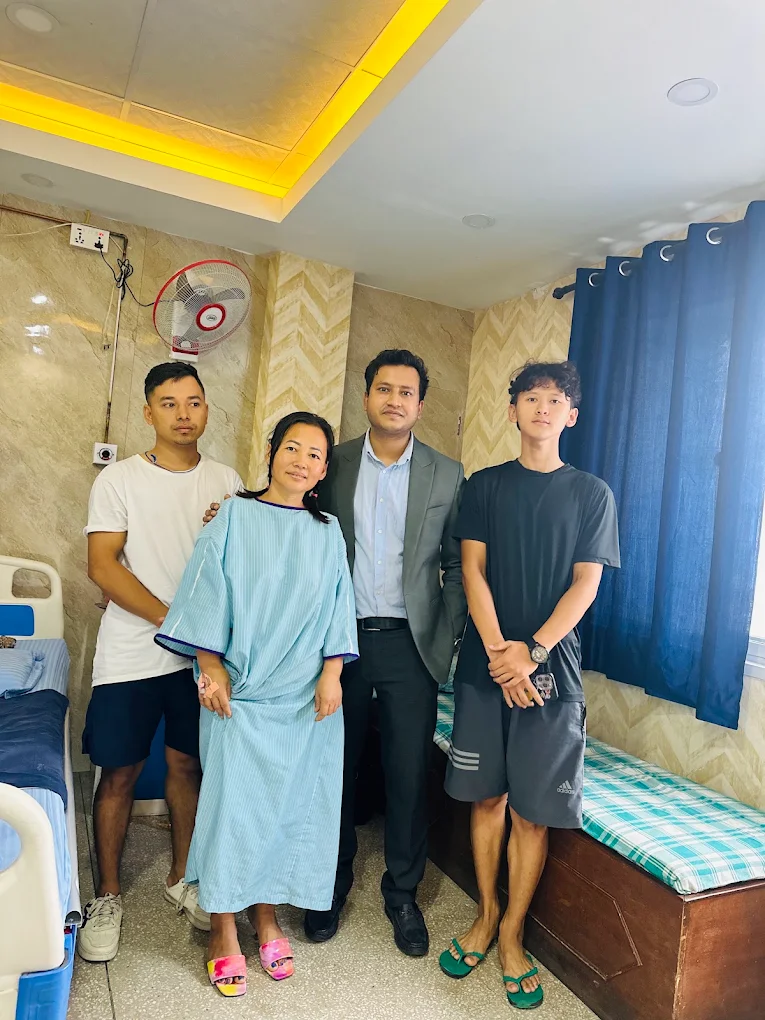
Infertility is defined as the inability to achieve a clinical pregnancy after 12 months of regular, unprotected intercourse (or after 6 months for women aged 35 and older). Successful management begins with a systematic, evidence-based diagnostic evaluation.
Diagnostic Evaluation Process
-
Ovarian Reserve Assessment: Hormonal testing including Serum AMH, Day 2/3 Follicle-Stimulating Hormone (FSH), Estradiol, and baseline transvaginal ultrasound for Antral Follicle Count (AFC).
-
Tubal and Uterine Cavity Patency: Hysterosalpingography (HSG) or combined Diagnostic Laparoscopy and Chromopertubation (dye test) to identify tubal blockages, hydrosalpinx, or pelvic peritubal adhesions.
-
Hysteroscopic Cavity Assessment: Direct visualization of the endometrial cavity to detect and correct endometrial polyps, submucosal fibroids, intrauterine synechiae (Asherman syndrome), or uterine septa.
-
Male Factor Assessment: Comprehensive semen analysis in accordance with World Health Organization (WHO) criteria to assess sperm count, motility, and morphology.
Personalized Treatment Pathways
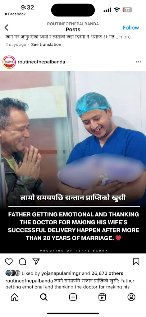
Based on diagnostic findings, Dr. Shah outlines structured treatment plans:
-
Ovulation Induction (OI) and Timed Intercourse: Tailored medication protocols for anovulatory cycles, often related to Polycystic Ovary Syndrome (PCOS), monitored via serial transvaginal follicular ultrasound scans.
-
Intrauterine Insemination (IUI): Preparation and placement of washed, motile sperm into the uterine cavity around the time of ovulation for couples with mild male factor subfertility, unexplained infertility, or cervical factors.
-
Advanced Reproductive Technology (ART / IVF-ICSI Counseling): Guiding couples through In Vitro Fertilization and Intracytoplasmic Sperm Injection pathways when severe male-factor subfertility, bilateral tubal obstruction, advanced maternal age, or long-standing unexplained infertility is present.
6. Laparoscopic Surgery vs. Traditional Open Surgery
Minimally invasive gynecological procedures have largely replaced conventional open laparotomy for many benign pelvic conditions. The table below summarizes key differences:
| Parameter | Advanced Laparoscopic Surgery | Traditional Open Laparotomy |
| Incision Size | 3 to 4 small keyhole incisions (5–10 mm) | One large abdominal incision (10–15+ cm) |
| Postoperative Pain | Mild to moderate; lower requirement for high-dose opioid analgesics | Moderate to severe due to abdominal wall muscle cutting |
| Hospital Stay | Typically 24 to 48 hours | Typically 4 to 7 days |
| Full Recovery Time | 1 to 2 weeks for most daily activities | 6 to 8 weeks for physical recovery |
| Intraoperative Blood Loss | Minimal, aided by high-definition visualization and precise vessel sealing | Higher, with wider exposure requirements |
| Risk of Adhesion Formation | Lower risk of postoperative peritoneal adhesions | Higher risk due to open air exposure and tissue handling |
| Cosmetic Result | Small, faint scars that fade over time | A prominent linear abdominal scar |
Note: While laparoscopy offers clear recovery advantages, the surgical approach must be individualized based on tumor size, suspicion of malignancy, previous abdominal surgeries, and underlying medical conditions.
7. What Patients Should Expect: From Consultation to Recovery
Step 1: Initial Outpatient Consultation
Patients undergo a detailed medical history review, physical examination, and pelvic ultrasonography. Prior laboratory reports, imaging, and surgical notes are evaluated to establish an accurate diagnosis.
Step 2: Pre-Operative Planning and Anesthesia Clearance
If surgery is indicated, routine pre-anesthesia clearance tests (complete blood counts, coagulation profiles, renal and liver function, viral screening, ECG, chest X-ray) are completed to ensure safety.
Step 3: Day of Surgery
Under general anesthesia, the procedure is completed using the Karl Storz 3D 4K RUBINA system. Small port sites are closed with fine subcuticular sutures that minimize visible scarring.
Step 4: Postoperative Care and Discharge
Patients are encouraged to mobilize gently within 6 to 12 hours after surgery to reduce the risk of deep vein thrombosis and aid bowel motility. Most patients return home within 24 to 48 hours with clear pain management and wound care instructions.
8. Frequently Asked Questions
What are the main advantages of 3D 4K laparoscopic surgery over traditional open surgery?
Laparoscopic surgery uses small 5 mm to 10 mm keyhole incisions instead of a large abdominal cut. The Karl Storz 3D 4K system provides magnified depth perception, reducing blood loss, tissue trauma, and postoperative pain. As a result, patients typically return home within 24 to 48 hours and resume normal daily activities within one to two weeks.
Can large uterine fibroids be safely removed laparoscopically?
Yes, many large and multiple uterine fibroids can be removed laparoscopically (laparoscopic myomectomy) by an experienced surgeon. The tissue is excised, the uterine wall is reconstructed with multi-layered sutures, and the fibroid tissue is removed through minimally invasive techniques. Suitability depends on the fibroid's location, number, and overall uterine size.
Will laparoscopic surgery for ovarian cysts decrease my fertility?
When performed with careful microsurgical techniques, laparoscopic cystectomy is designed to preserve fertility. By gently enucleating the cyst wall and minimizing excessive electrocautery near healthy ovarian tissue, the ovarian reserve is protected, helping safeguard future reproductive potential.
Where does Dr. Rajan Shah consult and perform surgeries in Nepal?
Dr. Rajan Shah conducts daily clinical consultations, outpatient evaluations, and advanced laparoscopic surgical procedures at Everest Hospital Pvt. Ltd., situated in New Baneshwor, Kathmandu, Nepal.
When should a couple seek an infertility evaluation?
Couples should seek an evaluation after 12 months of regular, unprotected intercourse without pregnancy if the female partner is under 35 years of age. If the female partner is 35 or older, or if there is a known history of irregular cycles, severe pelvic pain, endometriosis, or previous pelvic surgery, an evaluation is recommended after 6 months.
9. Key Takeaways and Clinical Perspective
-
Specialized Surgical Care: Dr. Rajan Shah has completed over 5,500 gynecological laparoscopic procedures, focusing on organ-preserving, minimally invasive solutions.
-
Modern Surgical Equipment: The Karl Storz 3D 4K RUBINA with ICG technology provides high-definition anatomical visualization, improving precision during complex pelvic operations.
-
Reproductive Focus: Surgical plans are structured around fertility preservation, combining diagnostic workups with corrective microsurgery and assisted reproductive techniques.
-
Central Practice Location: Based at Everest Hospital, New Baneshwor, Kathmandu, providing accessible, evidence-based care for patients throughout Nepal.
10. Schedule an Appointment
If you are experiencing symptoms of uterine fibroids, ovarian cysts, persistent pelvic pain, or are seeking a structured fertility evaluation, a comprehensive medical assessment is the first step.
Schedule a Consultation at Everest Hospital:
Visit the outpatient department at Everest Hospital Pvt. Ltd., New Baneshwor, Kathmandu, or book a clinical consultation at Everest Hospital to discuss your diagnosis and personalized treatment options with Dr. Rajan Shah.